 HOT
 BPAS-240 · 2026-11-17 · +86 21 5748 2007
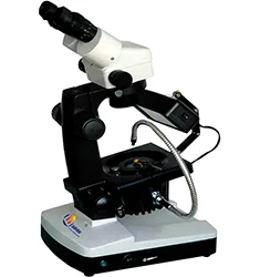
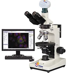
BPAS-240 Metallurgical Microscope
Metallurgical Microscope
모델BPAS-240
- 变倍范围连续变倍7X~45X(可扩展到50X)
- 工作距离95mm(可扩展至115mm);大视场广角目镜WF10×/23mm
- 可调瞳距范围55~75mm
- 调焦行程125mm
By submitting you agree to our privacy policy.
01
제품 개요
BPAS-240立体珠宝显微镜能够提供高清晰和高分辨率的立体图像。
BPAS-240立体宝石显微镜技术参数:
变倍范围:连续变倍7X~45X(可扩展到50X);
工作距离:95mm(可扩展至115mm);
大视场广角目镜WF10×/23mm;
可调瞳距范围:55~75mm;
调焦行程:125mm;
选配件:辅助物镜、目镜, 辅助物镜0.75×工作距离105mm,1.5×工作距离47mm,2×工作距离33mm。
技术优势:
灯管的连接方式更便于调节照明角度。调焦行程125mm,可对大小不同的宝玉石及样品进行观察。钢丝宝石夹钢性适中夹持牢固、方便,由于减少宝石腰部的光线遮挡,可以对宝石多方面地观察。工作台两边有宝石夹的安装孔,从而可以调节观察眼点的高度和角度,使操作更加方便舒适。
光源配置:
光源配置合理。附件宝石夹提供钢丝夹和平口夹,可供用户选用。偏光器使用能更好地观察到宝石内外结构的晶体特征,满足不同的使用要求。
BPAS-240 Metallurgical Microscope supplied by CHWES. 4 key specifications available on request.
02
사양
| 变倍范围 | 连续变倍7X~45X(可扩展到50X) |
|---|---|
| 工作距离 | 95mm(可扩展至115mm);大视场广角目镜WF10×/23mm |
| 可调瞳距范围 | 55~75mm |
| 调焦行程 | 125mm |
* Generated by rule engine over the product database; for pre-selection only.
03
측정 원리
The metallurgical microscope uses an infinity-corrected optical path with reflected Köhler illumination. Light passes the collector, field stop and aperture stop, is deflected by a semi-transparent mirror through the objective onto the specimen, and returns through the same objective to the eyepiece or camera.
- Bright field for routine microstructure, inclusions and decarburised layers.
- Dark field collects scattered light to reveal grain boundaries, cracks and non-metallic inclusions.
- Polarised light identifies anisotropic phases, spheroidised structures and coating stress.
- DIC converts tiny height differences into contrast, ideal for polished, unetched samples.
Objectives of 5× to 100× are combined with 10× eyepieces. Higher numerical aperture gives better resolution but shallower depth of field.
04
Process animation
- Sampling and preparation: sectioning, mounting, grinding, polishing, etching.
- Illumination and focus: select BF/DF/POL/DIC and focus.
- Image capture: trinocular port, 1× camera adapter, white balance.
- Scale calibration: calibrate µm per pixel with a stage micrometer.
- Measurement: grain size, case depth, coating thickness, inclusion rating.
05
Calculation & formulas
1. System magnification: M = objective × eyepiece × adapter, e.g. 40× / 10× / 1× = 400×.
2. Resolution: d = 0.61λ/NA (λ≈550 nm). A 40× objective with NA 0.65 resolves 0.52 µm; an NA 0.95 dry objective reaches 0.35 µm.
3. Depth of field scales with λ/NA² — higher NA, shallower depth; use low NA for fracture surfaces.
4. Grain size (GB/T 6394): at 100×, grains N per 645 mm² field follow N = 2G−1; counting 128 grains gives G = log2128 + 1 = 8. Intercept method: mean intercept L̄ = line length ÷ intercepts, G = −6.6437 lg L̄ (mm, 100×).
5. Effective case depth: distance to 550 HV1, fitted from a micro-hardness traverse and averaged over three positions.
06
규격
| Standard | Title | Scope |
|---|---|---|
| GB/T 13298-2015 | Metallic materials — examination of microstructure | Microstructure rating |
| GB/T 6394-2017 | Determination of average grain size | Grain size |
| ASTM E3-11 | Guide for metallographic specimen preparation | Preparation |
| ASTM E112 | Determining average grain size | Grain size |
| ISO 643 | Steels — micrographic determination of grain size | Grain size |
Export projects often require dual GB and ASTM reports; the software ships both rating charts.
07
선택 가이드
1. Surface or cross-section: stereo microscopes (HX-ZG7, XSZ) for fracture surfaces, solder joints and coatings; upright or inverted metallurgical microscopes (HX-4, HX-G series) for microstructure, case depth and inclusions.
2. Upright or inverted: upright for small specimens that are flipped often; inverted (HX-G3A/G4C/G5A) for large, heavy parts such as gears, shafts and tube sections that go straight onto the stage without re-mounting.
3. Optics set the ceiling: plan achromats cover routine rating; semi-apochromats or apochromats with a research stand (HX-G5A) are needed for 1000× precipitates and brightfield/darkfield/polarised switching.
4. Three levels of digitalisation: a 5 MP camera for images; camera plus analysis software (grain size, case depth, inclusions, porosity) for automatic rating and reports; motorised stage with scanning and stitching for unattended batches.
5. Do not forget accessories: polariser, darkfield, DIC, and a microhardness interface.
6. Room: an anti-vibration bench at 20±3 °C for research stands; keep preparation and microscopy rooms separate to avoid abrasive dust.
| Series | Type | Typical task | Recommended setup |
|---|---|---|---|
| HX-4A / HX-4B | Upright metallurgical | Routine structure, case depth | 5×/10×/40× + 5 MP camera |
| HX-G2A / G2B | Inverted | Batch samples, no re-mounting | Brightfield + simple polariser |
| HX-G3A | Trinocular inverted | Case depth, rating, reports | Camera + analysis software |
| HX-G4C / G5A | Research inverted | Darkfield/polarised/DIC | Semi-apochromat + motorised stage |
| HX-ZG7 / XSZ-146 | Stereo | Fracture, solder, low-mag locating | Zoom + ring light |
Common misconceptions: higher magnification is not better (numerical aperture limits resolution, 100× dry is the practical ceiling); ignoring stage travel so large samples do not fit; buying the stand without software and still rating by eye, which kills repeatability.
08
시험 방법
Preparation. Section perpendicular to the feature of interest. Keep hot-mounting below 150 °C for aluminium and quenched parts, or use cold mounting. Grind 320→600→800→1200, removing previous scratches at each step; polish with 3 µm then 1 µm diamond. Etchants: 4% nital for carbon steel (3–8 s), aqua regia or electrolytic for stainless, Keller for aluminium.
Examination. Locate a representative field at 5×/10×, then switch to higher power. Rate grain size to GB/T 6394 by comparison or intercept. Measure case depth at 500× with an ocular scale or calibrated software, averaging five readings.
Calibration. Verify µm/pixel daily with a stage micrometer, check against a certified specimen quarterly, record three fields per sample and archive images with scale bars.
Pitfalls. Over-etching widens boundaries and fakes decarburisation; residual scratches can be mistaken for cracks; rounded mounting edges inflate case depth. Keep an unetched specimen for coating and crack review.
09
Industry applications
- Steel and heat treatment: decarburised depth, carburised/nitrided case depth, grain size (GB/T 6394), non-metallic inclusions (GB/T 10561), banding and Widmanstätten rating.
- Non-ferrous metals: aluminium grain and recrystallisation, alpha/beta ratio in brass, twinning and corrosion morphology in magnesium.
- Welding and brazing: penetration, HAZ width, porosity and cracks, intermetallic thickness at dissimilar joints.
- Additive manufacturing: porosity, lack-of-fusion defects, melt-pool morphology in SLM parts, porosity repeatability within 0.2%.
- Bearings and gears: martensite rating, retained austenite, carbide network and banding, grinding burn and white layer.
- Electronics: IMC thickness, plating cross-sections, PCB micro-sections, gold wire ball bonds (stereo plus metallurgical).
- Powders and ceramics: particle morphology and size, sintered porosity, coating cross-section thickness.
- Failure analysis: fracture surface macro and micro morphology, corrosion products, crack path and origin, with stereo microscopy for low-magnification locating.
10
Price and lead time for this model
동일 시리즈
전체 보기→Microscopes
HOT HOT
 HOT