 HOT
 AMM-17 · 2026-11-17 · +86 21 5748 2007
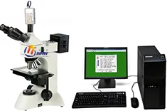
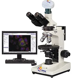
AMM-17 Metallurgical Microscope
Metallurgical Microscope
ModellAMM-17
- 正置明暗视场透反射金相显微镜数码正置明暗视场透反射金相显微 / 视频图像正置明暗视场透反射金相显微镜 / 图像处理正置明暗视场透反射金相显微镜 / 数据处理正置明暗视场透反射金相显微镜
- 1、目镜标配:大视野 WF10X(视场数Φ22mm)
- 2、物镜无限远长工作距离平场消色差明/暗视场物镜(无盖玻片)
- 3、选购物镜PL L50X/0.70B.D 工作距离:2.50mm
By submitting you agree to our privacy policy.
01
Überblick
AMM-17系列正置明暗视场透反射金相显微镜是一种多用途工业检验用光学仪器,配置明/暗场物镜、大视野目镜与偏光观察装置,透反射照明采用“柯勒”照明系统,视场清晰。可用于半导体硅晶片、LCD基板、电路板、固体粉未及其它各种透明或不透明工业试样的检验,是生物学、金属学、矿物学、精密工程学、电子工程学等研究的理想仪器.
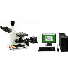
AMM-17D数码正置明暗视场透反射金相显微镜是将传统的光学显微镜与数码相机通过光电转换有机的结合在一起,不仅可以在目镜上作显微观察,还能在数码相机显示屏幕上观察实时动态图像,并能将所需要的图片进行编辑、保存和打印。
AMM-17P视频图像正置明暗视场透反射金相显微镜是将传统的光学显微镜与图像处理通过光电转换有机的结合在一起,不仅可以在目镜上作显微观察,还能在视频显示屏幕上观察实时动态图像,同时还可以对金相组织结构进行直观明了的观察等。
AMM-17T图像处理正置明暗视场透反射金相显微镜是将传统的光学显微镜与计算机通过光电转换有机的结合在一起,不仅可以在目镜上作显微观察,还能在计算机显示屏幕上观察实时动态图像,并能将所需要的图片进行编辑、保存和打印。
AMM-17ST数据处理正置明暗视场透反射金相显微镜是将传统的光学显微镜与计算机通过光电转换有机的结合在一起,不仅可以在目镜上作显微观察,还能在计算机显示屏幕上观察实时动态图像,并能将所需要的图片进行编辑、保存和打印,同时还可以对金相组织结构进行数据处理分析,对照金相模块自动评级等。
二、AMM-17系列正置明暗视场透反射金相显微镜性能特点:
1、采用无限远光学系统及模块化功能设计。
2、长工作距离明/暗视场平场物镜,视场大而清晰。
3、广角平场目镜:视场直径Ф22mm。
4、粗微动同轴调焦机构,粗动松紧可调,带限位锁紧装置,微动格值:2μm.
5、内置式可切换偏光观察装置,起偏器可360°旋转。
6、上光源采用翻盖式12V50W卤素灯箱,更换灯炮安全便捷,下光源采用12V30W卤素灯,加装散热片。
9、三目镜筒可自由切换目视观察与显微摄影,摄影时可100%通光,适合暗视场显微图像拍摄。
三、AMM-17系列正置明暗视场透反射金相显微镜技术参数:
型号MC006
AMM-17
AMM-17D
AMM-17P
AMM-17T
AMM-17ST
正置明暗视场透反射金相显微镜
数码正置明暗视场透反射金相显微
视频图像正置明暗视场透反射金相显微镜
图像处理正置明暗视场透反射金相显微镜
数据处理正置明暗视场透反射金相显微镜
1、目镜
标配:大视野 WF10X(视场数Φ22mm)
选购:分划目镜10X(视场数Φ22mm) 格值0.10mm/格
2、物镜
无限远长工作距离平场消色差明/暗视场物镜(无盖玻片)
PL L5X/0.12 B.D 工作距离(Work distance):9.7 mm
PL L10X/0.25 B.D 工作距离(Work distance):9.3 mm
PL L20X/0.40 B.D 工作距离(Work distance):7.2 mm
PL L40X/0.60 B.D 工作距离(Work distance):3.0 mm
3、选购物镜
PL L50X/0.70B.D 工作距离:2.50mm
PL L60X/0.70 B.D 工作距离:1.90 mm
PL L80X/0.80B.D 工作距离:1.25mm
PL L100X/0.85B.D 工作距离:0.22mm
4、目镜筒
三目镜,倾斜30度,(内置检偏振片,可进行切换)
5、落射照明系统
12V 50W卤素灯,亮度与灯炮位置可调
内置视场光阑、孔径光阑、滤色片转换装置,推拉式检偏器与起偏器
6、调焦机构
粗微动同轴调焦, 微动格值:2μm,带锁紧和限位装置
7、转换器
五孔(内向式滚珠内定位)
8、载物台
双层机械移动式(尺寸:210mmX140mm,移动范围:75mmX50mm)
9、透射照明系统
阿贝聚光镜 NA.1.25 可上下升降
蓝滤色片和磨砂玻璃,选购:黄滤色片、绿滤色片。
集光器,卤素灯照明适用(内置视场光栏)
12V/30W卤素灯,亮度可调
10、配备系统
标准主机1套
主机+数码成像系统1套
主机+视频图像处理系统1套
主机+图像处理系统1套
主机+数据处理系统1套
AMM-17 Metallurgical Microscope supplied by CHWES. 11 key specifications available on request.
02
Technische Daten
| 正置明暗视场透反射金相显微镜 | 数码正置明暗视场透反射金相显微 / 视频图像正置明暗视场透反射金相显微镜 / 图像处理正置明暗视场透反射金相显微镜 / 数据处理正置明暗视场透反射金相显微镜 |
|---|---|
| 1、目镜 | 标配:大视野 WF10X(视场数Φ22mm) |
| 2、物镜 | 无限远长工作距离平场消色差明/暗视场物镜(无盖玻片) |
| 3、选购物镜 | PL L50X/0.70B.D 工作距离:2.50mm |
| 4、目镜筒 | 三目镜,倾斜30度,(内置检偏振片,可进行切换) |
| 5、落射照明系统 | 12V 50W卤素灯,亮度与灯炮位置可调 |
| 6、调焦机构 | 粗微动同轴调焦, 微动格值:2μm,带锁紧和限位装置 |
| 7、转换器 | 五孔(内向式滚珠内定位) |
| 8、载物台 | 双层机械移动式(尺寸:210mmX140mm,移动范围:75mmX50mm) |
| 9、透射照明系统 | 阿贝聚光镜 NA.1.25 可上下升降 |
| 10、配备系统 | 标准主机1套 / 主机+数码成像系统1套 / 主机+视频图像处理系统1套 / 主机+图像处理系统1套 / 主机+数据处理系统1套 |
* Generated by rule engine over the product database; for pre-selection only.
03
Messprinzip
The metallurgical microscope uses an infinity-corrected optical path with reflected Köhler illumination. Light passes the collector, field stop and aperture stop, is deflected by a semi-transparent mirror through the objective onto the specimen, and returns through the same objective to the eyepiece or camera.
- Bright field for routine microstructure, inclusions and decarburised layers.
- Dark field collects scattered light to reveal grain boundaries, cracks and non-metallic inclusions.
- Polarised light identifies anisotropic phases, spheroidised structures and coating stress.
- DIC converts tiny height differences into contrast, ideal for polished, unetched samples.
Objectives of 5× to 100× are combined with 10× eyepieces. Higher numerical aperture gives better resolution but shallower depth of field.
04
Process animation
- Sampling and preparation: sectioning, mounting, grinding, polishing, etching.
- Illumination and focus: select BF/DF/POL/DIC and focus.
- Image capture: trinocular port, 1× camera adapter, white balance.
- Scale calibration: calibrate µm per pixel with a stage micrometer.
- Measurement: grain size, case depth, coating thickness, inclusion rating.
05
Calculation & formulas
1. System magnification: M = objective × eyepiece × adapter, e.g. 40× / 10× / 1× = 400×.
2. Resolution: d = 0.61λ/NA (λ≈550 nm). A 40× objective with NA 0.65 resolves 0.52 µm; an NA 0.95 dry objective reaches 0.35 µm.
3. Depth of field scales with λ/NA² — higher NA, shallower depth; use low NA for fracture surfaces.
4. Grain size (GB/T 6394): at 100×, grains N per 645 mm² field follow N = 2G−1; counting 128 grains gives G = log2128 + 1 = 8. Intercept method: mean intercept L̄ = line length ÷ intercepts, G = −6.6437 lg L̄ (mm, 100×).
5. Effective case depth: distance to 550 HV1, fitted from a micro-hardness traverse and averaged over three positions.
06
Normen
| Standard | Title | Scope |
|---|---|---|
| GB/T 13298-2015 | Metallic materials — examination of microstructure | Microstructure rating |
| GB/T 6394-2017 | Determination of average grain size | Grain size |
| ASTM E3-11 | Guide for metallographic specimen preparation | Preparation |
| ASTM E112 | Determining average grain size | Grain size |
| ISO 643 | Steels — micrographic determination of grain size | Grain size |
Export projects often require dual GB and ASTM reports; the software ships both rating charts.
07
Auswahl
1. Surface or cross-section: stereo microscopes (HX-ZG7, XSZ) for fracture surfaces, solder joints and coatings; upright or inverted metallurgical microscopes (HX-4, HX-G series) for microstructure, case depth and inclusions.
2. Upright or inverted: upright for small specimens that are flipped often; inverted (HX-G3A/G4C/G5A) for large, heavy parts such as gears, shafts and tube sections that go straight onto the stage without re-mounting.
3. Optics set the ceiling: plan achromats cover routine rating; semi-apochromats or apochromats with a research stand (HX-G5A) are needed for 1000× precipitates and brightfield/darkfield/polarised switching.
4. Three levels of digitalisation: a 5 MP camera for images; camera plus analysis software (grain size, case depth, inclusions, porosity) for automatic rating and reports; motorised stage with scanning and stitching for unattended batches.
5. Do not forget accessories: polariser, darkfield, DIC, and a microhardness interface.
6. Room: an anti-vibration bench at 20±3 °C for research stands; keep preparation and microscopy rooms separate to avoid abrasive dust.
| Series | Type | Typical task | Recommended setup |
|---|---|---|---|
| HX-4A / HX-4B | Upright metallurgical | Routine structure, case depth | 5×/10×/40× + 5 MP camera |
| HX-G2A / G2B | Inverted | Batch samples, no re-mounting | Brightfield + simple polariser |
| HX-G3A | Trinocular inverted | Case depth, rating, reports | Camera + analysis software |
| HX-G4C / G5A | Research inverted | Darkfield/polarised/DIC | Semi-apochromat + motorised stage |
| HX-ZG7 / XSZ-146 | Stereo | Fracture, solder, low-mag locating | Zoom + ring light |
Common misconceptions: higher magnification is not better (numerical aperture limits resolution, 100× dry is the practical ceiling); ignoring stage travel so large samples do not fit; buying the stand without software and still rating by eye, which kills repeatability.
08
Prüfverfahren
Preparation. Section perpendicular to the feature of interest. Keep hot-mounting below 150 °C for aluminium and quenched parts, or use cold mounting. Grind 320→600→800→1200, removing previous scratches at each step; polish with 3 µm then 1 µm diamond. Etchants: 4% nital for carbon steel (3–8 s), aqua regia or electrolytic for stainless, Keller for aluminium.
Examination. Locate a representative field at 5×/10×, then switch to higher power. Rate grain size to GB/T 6394 by comparison or intercept. Measure case depth at 500× with an ocular scale or calibrated software, averaging five readings.
Calibration. Verify µm/pixel daily with a stage micrometer, check against a certified specimen quarterly, record three fields per sample and archive images with scale bars.
Pitfalls. Over-etching widens boundaries and fakes decarburisation; residual scratches can be mistaken for cracks; rounded mounting edges inflate case depth. Keep an unetched specimen for coating and crack review.
09
Industry applications
- Steel and heat treatment: decarburised depth, carburised/nitrided case depth, grain size (GB/T 6394), non-metallic inclusions (GB/T 10561), banding and Widmanstätten rating.
- Non-ferrous metals: aluminium grain and recrystallisation, alpha/beta ratio in brass, twinning and corrosion morphology in magnesium.
- Welding and brazing: penetration, HAZ width, porosity and cracks, intermetallic thickness at dissimilar joints.
- Additive manufacturing: porosity, lack-of-fusion defects, melt-pool morphology in SLM parts, porosity repeatability within 0.2%.
- Bearings and gears: martensite rating, retained austenite, carbide network and banding, grinding burn and white layer.
- Electronics: IMC thickness, plating cross-sections, PCB micro-sections, gold wire ball bonds (stereo plus metallurgical).
- Powders and ceramics: particle morphology and size, sintered porosity, coating cross-section thickness.
- Failure analysis: fracture surface macro and micro morphology, corrosion products, crack path and origin, with stereo microscopy for low-magnification locating.
10
Price and lead time for this model
Ähnliche Modelle
Alle→Microscopes
HOT HOT
 HOT